
Instruct-EMBL Annual Report Highlights and New Technologies
The European Molecular Biology Laboratory (EMBL) is Europe’s flagship laboratory for the life sciences. EMBL is an intergovernmental organisation supported by 28 countries: Instruct Centre EMBL Grenoble, Instruct Centre EMBL Hamburg, and Instruct Centre EMBL Heidelberg. Find out more about Instruct-EMBL and apply to access technologies here.
Contacts: Kristina Djinovic Carugo, Jose A. Marquez, Matthias Wilmanns, Thomas Schneider, Christoph Müller, Simone Mattei
Instruct Centre EMBL Grenoble hosts one of the largest platforms for high-throughput crystallisation screening in Europe. The HTX lab offers automated protein-to-structure and fragment screening crystallography pipelines, including screening and structural characterisation of ligand-target complexes.
Instruct Centre EMBL Hamburg focuses on state-of-the-art structural biology methods using synchrotron radiation. It operates three beamlines on the PETRA III high-brilliance synchrotron. The Sample Preparation and Characterisation Facility (SPC) enables access to sample preparation and molecular biophysics characterisation of macromolecular complexes.
Instruct Centre EMBL Heidelberg specialises in high-resolution cryogenic electron microscopy (cryo-EM) of biological samples, supporting users for single-particle and tomography projects.
Technological Advances in 2022
Instruct Centre EMBL Grenoble - Two in one: combining MASSIF-1 and CrystalDirect
Jointly operated by EMBL Grenoble and the European Synchrotron Radiation Facility (ESRF), MASSIF-1, is the only fully automated beamline dedicated to structural biology worldwide. Teams from EMBL and ESRF have further improved its workflows and modalities in 2022 with the integration of a CrystalDirectTM harvester. Macromolecular crystallography is one of the techniques used to determine the 3D structures of macromolecules, like proteins, by shooting X-rays at regularly structured arrays of molecules, packed together in a crystal. The whole process involves several manual steps, including growing and harvesting the crystal. The CrystalDirectTM technology consists of an extremely precise robot that streamlines the step of crystal sample harvesting and preparation. It automatically harvests and cryo-cools the crystal. The CrystalDirectTM robotic harvester has been installed into the experimental hutch of MASSIF-1 allowing the beamline to harvest crystals and collect data simultaneously. The integration of the CrystalDirect technology into MASSIF-1 improves the beamline capacities and expands the service portfolio of the HTX lab. Now EMBL and ESRF are jointly offering a completely automated ‘plate-to-beam’ service, remotely accessible to users via the HTX lab.
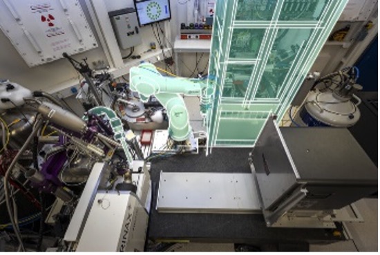
The CrystalDirect harvester (top right) integrated into the experimental hutch of the MASSIF-1 beamline, together with two pieces of equipment for sample transfer and humidity control. Credits: Stuart Ingham/EMBL, Isabel Romero Calvo/EMBL
Instruct Centre EMBL Hamburg - expansion of eSPC service
During 2022, the SPC facility at Hamburg has seen an important expansion of its eSPC services. The eSPC, is a freely available online platform for analysing molecular biophysics data from a range of experimental techniques: Differential scanning fluorimetry, Fluorescence quenching and Microscale thermophoresis. The team expanded the analysis to Mass Photometry and Dynamic light scattering with the release of the PhotoMol and Raynals apps.
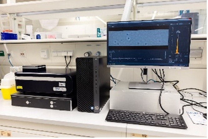
MassPhotometer (Refeyn) at the SPC facility, data analysis for this and equivalent instruments can be performed remotely.
Mass photometry is used to assess reconstitution efficiency, and oligomerisation and size distribution profile of samples. The main objective of PhotoMol is to estimate the masses of different species in a sample by analysing mass photometry data. The PhotoMol pipeline consists of three steps. First, the user loads an input file that contains the frequency of the observed masses (or contrasts). Pre-processing is first performed by selecting a bin width and a window range to build the histogram. Second, the user defines the number of Gaussians (species) present in the distribution and a truncated multi-Gaussian fit is executed. Finally, publication-grade figures can be downloaded together with information about the fitted parameters. Dynamic light scattering (DLS) is routinely employed to assess the aggregation behaviour, and homogeneity of samples containing microscopic particles in suspension or solubilised polymers. To improve its service offering, the SPC developed Raynals, a user-friendly software for the analysis of single-angle DLS data that uses the Tikhonov-Phillips regularisation. DLS data can be easily misinterpreted and the simulation tools available in Raynals allow understanding of the limitations of the measurement and its resolution. It has been designed as a tool to address quality control of biological samples, during sample preparation and optimisation, and it helps in the detection of aggregates showing the influence of large particles. Lastly, Raynals provides flexibility in the way the data is presented, allowing exporting publication-quality figures.
Instruct Centre EMBL Heidelberg - cryo-CLEM now available to users.
In 2022 Heidelberg added Cryo Correlative Light and Electron Microscopy (CLEM) to its catalogue of services. Correlative microscopy utilises complementary visual techniques such as light/fluorescence microscopy and electron microscopy that allow the experimenter to navigate large cellular volumes, to capture significant proportions of a population of cells, to identify features of interest, and to then acquire high-resolution snapshots that represent cellular events. After cryofixation by plunge or high pressure freezing, samples can be imaged in a cryo-confocal microscope (Zeiss LSM 900 Airyscan2 and Leica cryo-Stellaris 5) and subsequently processed further in a cryo-FIB-SEM (ThermoScientific Aquilos 2 and Zeiss Crossbeam 550). FIB-milling of target areas allows the creation of lamellae that can be subsequently imaged by tomography in a 300 kV ThermoScientific Titan Krios G4 TEM.
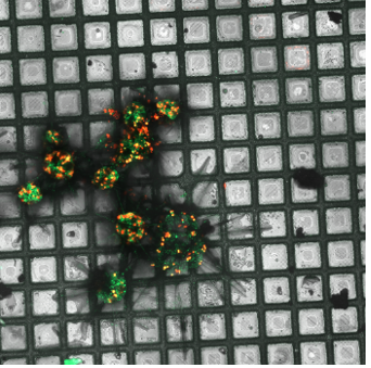
Maximum intensity projection of Acantharia (plankton), plunge frozen and imaged with Zeiss LSM 900 Airyscan2. Green: Acantharia; red: chlorophyll of symbionts (microalgae). Credits: Anna Steyer/EMBL
EMBL Structural Biology Activities
- Olivier Duss (Group Leader at EMBL Heidelberg) was awarded an Instruct-ERIC Pilot R&D 2022 grant. His project will examine “In vivo single-molecule imaging of in vitro reconstituted macromolecular machineries”. He plans to combine the advantages of both in vitro and in vivo approaches by first reconstituting complicated macromolecular systems in vitro and then electroporating them into live E.coli cells for real-time in vivo imaging of complicated processes in physiological native context. He hopes for the first time be able to track directly the process of translation in vivo at single amino-acid resolution.
- EMBL Hamburg welcomed Leonardo Alonso (University of Buenos Aires) as part of a Res Infra Staff Exchanges Call. He joined Maria Garcia’s group to continue his work on Deamidation of SARS-CoV-2 spike protein and its role in immune evasion. This successful exchange has already led to further collaborations and visits. This project is a continuation of work, resulting in this publication.
- Two early career researchers from EMBL Heidelberg were invited as speakers to the Instruct Biennial 2022. Agata Misiaszek, a pre-doc in Christoph Müller’s group, presented her work on “Cryo-EM structures of human RNA polymerase I”. While Liang Xue, a post-doc in Julia Mahamid’s group, spoke on his work on “Visualising translation dynamics and landscapes in unprecedented detail with cryo-ET and subtomo analysis”.
- EMBL Heidelberg welcomed Adria Alcaide Jimenez from IBMB- CSIC as part of Instruct’s Internship programme. Adria joined Christoph Müller’s lab where he worked on high-resolution cryo-EM structures of virulent transcription complexes of Vibrio cholerae. His internship has already led to further collaboration, and an Instruct funded visit.
- The EMBL Imaging centre (IC) began operations in late 2021, with its official opening in June 2022. The IC has enjoyed a successful first year, welcoming a wide variety of users, including many from Instruct. As part of its opening it hosted the first EMBL Imaging Centre symposium. This introduced the ICs services, aims, and missions to the scientific community, celebrated advancements in imaging, and provided a sneak-peek into the cutting-edge technologies and advanced training it will make accessible to researchers all over the world. The speakers, including Nobel Laureates Stefan Hell, Richard Henderson, and W.E. Moerner, spoke on the rapid developments in imaging technologies that have led to notable biological and medical discoveries, the future trends in Cryo-EM, and challenges that would be faced.
Artistic visual summary of microscopy techniques available at the Imaging Centre. Credit: Isabel Romero Calvo/EMBL