18-Dec-2023
A new 200kV (scanning-)transmission electron microscope for life-science applications has been installed at Cryo-electron microscopy core facility (CEMCOF), part of the CEITEC facility in Instruct-CZ. The Glacios 2 (ThermoScientific) microscope will be available exclusively for cryo-electron microscopy, including efficient screening of cryo-EM samples, single particle data acquisition, cryo-electron tomography data acquisition, cryo-STEM imaging and tomography. The single particle cryo-EM data can be collected in fringe-free mode to maximise the efficiency of the data collection.

200kV (scanning-)transmission electron microscope: Glacios 2 (ThermoScientific) at CEITEC Instruct-CZ
The microscope is equipped with a Falcon 4i direct electron detector for TEM applications, a Ceta-D camera for electron diffraction tomography, and a set of bright-field, dark-field and high angle annular dark-field detectors (Panther STEM, ThermoScientific) for STEM imaging. Data acquisition is automated using SerialEM, EPU Multigrid and Tomo5 (including Tomo-Live for real-time tomogram reconstruction). The microscope at CEMCOF is the 200th Glacios installed worldwide.
A new focused ion beam scanning electron microscope (FIB/SEM) equipped is available for service at Cryo-electron microscopy and tomography core facility (CEMCOF) at CEITEC Instruct centre (Czech Republic). Helios Hydra V (ThermoScientific) is equipped with an immersion SEM column for high-resolution SEM imaging, FIB working on the principle of inductively-couple plasma with a four gas (Xe,Ar,O,N) source for efficient material ablation.
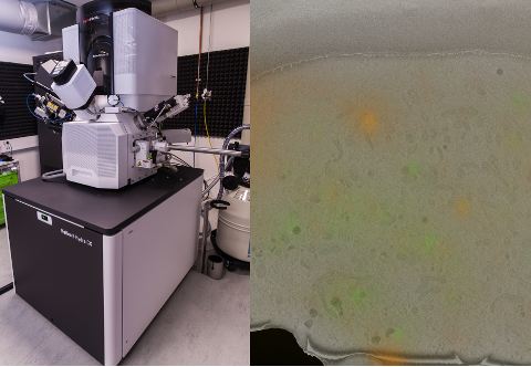
Correlative cryo-SEM and cryo-FM imaging of the pancreatic INS-1E cells (right). The cellular membrane (red) and mitochodria (green) were labeled dyes for live cell imaging. The data were collected using Helios Hydra V (left) equipped with the integrated flourescence microscope.
The available services will focus on the targeted lamella preparation using correlative light-electron microscopy and other correlative studies thanks to the wide-field fluorescence microscope integrated into the SEM chamber. In addition, Helios Hydra is available for FIB/SEM volume imaging under cryo- and room-temperatures conditions to support research focused on ultrastructural analysis of the whole cells and tissue regions.
Find out more about the CEITEC facility and access technologies here.
